
Table of Contents
- You Have a Report You Cannot Understand
- What the Doppler Machine Is Doing
- The Key Terms on Your Report
- Examples of Reassuring Findings and Reflux Findings
- What Happens After the Report
1. You Have a Report You Cannot Understand
Your Doppler scan is done. Now you are holding a report with phrases such as “reflux in the GSV”, “SFJ incompetence” and “reflux duration 4.2 seconds”. You may have been told to show it to your doctor, but you would still like to understand what it says.
Searching one term at a time can make the report more confusing. A definition rarely tells you what a finding means for your own symptoms.
In this guide, I explain the common terms and the questions they help answer. The report is useful information, but it needs to be read alongside your symptoms, medical history and an examination.
2. What the Doppler Machine Is Doing
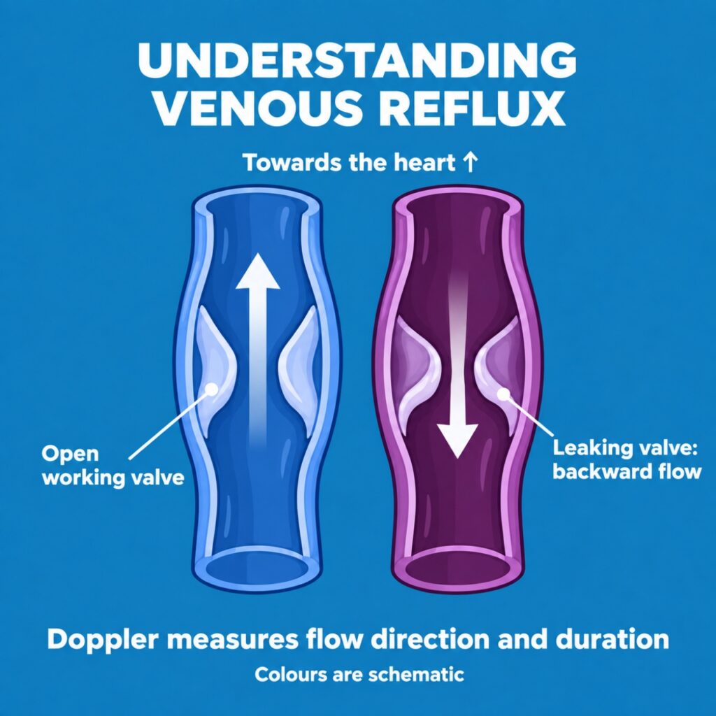
A venous duplex ultrasound uses sound waves to show the veins and assess blood flow. It does not use ionising radiation. The examination can look for blockage and check whether the valves that help blood return towards the heart are working properly.
The red and blue colours describe flow direction relative to the ultrasound probe. They do not, by themselves, mean artery versus vein, or healthy versus unhealthy blood flow. Probe position and the colour scale matter.
Think of the valves as small gates helping to limit backward flow. If the gates do not close effectively, blood can flow back and increase pressure in the veins. This backward flow is called reflux. It is a common finding in varicose veins.
The sonographer may press on a vein to check whether it compresses and squeeze your calf to assess flow. These are different parts of the examination.
For reflux testing, standing is preferred when feasible; a sitting or tilted position may be used if you cannot stand. A scan focused on suspected deep vein thrombosis and a detailed reflux study do not necessarily answer all the same questions.
3. The Key Terms on Your Report
GSV or Great Saphenous Vein
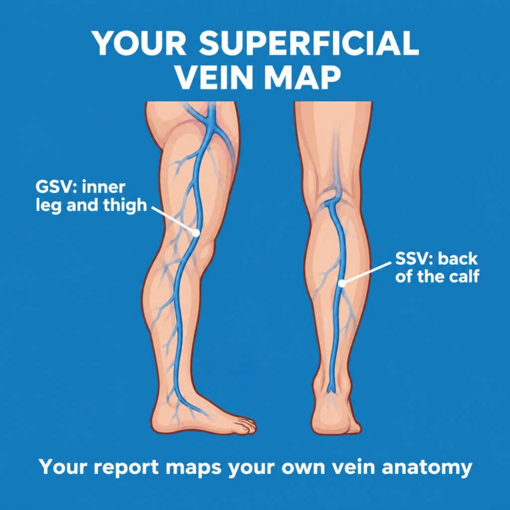
This is a major superficial vein running along the inner side of the leg and thigh towards the groin. “Superficial” means it belongs to the vein system nearer the skin; it does not mean the finding is unimportant.
The GSV can be a source of reflux contributing to visible varicose veins. Treating it may be part of the plan, but visible branches can also need treatment.
SSV or Small Saphenous Vein
This vein runs up the back of the calf. It usually joins a deep vein behind the knee, although its connections vary between people. That variation is one reason ultrasound mapping is useful before treatment.
SFJ or Saphenofemoral Junction
This is where the GSV joins the common femoral vein in the groin. “SFJ incompetence” describes reflux at this junction. It is a location in the vein network, not a description of the whole leg.
SPJ or Saphenopopliteal Junction
This is the usual junction between the SSV and the popliteal vein behind the knee. Your report may describe a different connection because the anatomy varies.
Reflux and Reflux Duration
Reflux duration is the length of time backward flow is recorded during a test manoeuvre, such as calf compression and release.
Common guideline thresholds are:
| Vein being assessed | Reversed flow used to define reflux |
| Superficial trunk veins such as the GSV and SSV | More than 0.5 seconds |
| Common femoral, femoral and popliteal veins | More than 1 second |
| Tibial, deep femoral and perforating veins | More than 0.5 seconds |
These values are interpreted with the test technique and the vein being examined. The threshold is not the same for every vein.
One duration should not be used to rank the overall severity of your disease or decide on treatment by itself. Symptoms, skin changes and the distribution of reflux also matter.
Perforator Incompetence
Perforators are connecting veins between the superficial and deep systems. An incompetent perforator can allow outward flow towards superficial veins. Its importance depends on the location, vein diameter and your symptoms or skin changes. It does not automatically need a separate procedure.
Deep Veins Patent and No DVT
Patent means open. DVT means deep vein thrombosis, a blood clot in a deep vein. “No DVT” means no clot was identified in the areas assessed at that examination; it is reassuring, but the scan must still be interpreted in its clinical context.
An open vein may still have reflux, so “patent” does not mean that all deep vein valves have been shown to work normally.
If there is ongoing concern about a clot, a doctor may advise repeat imaging or other assessment after an initially negative scan. Attend any follow-up you are given, particularly if symptoms persist or worsen.
4. Examples of Reassuring Findings and Reflux Findings
An illustrative report with reassuring findings: “No reflux identified in the assessed GSV or SSV segments. Deep veins patent. No evidence of DVT in the examined veins.”
This suggests that the scan has not identified reflux in those superficial veins or a clot in the examined deep veins. It does not establish that every possible cause of leg pain or swelling has been excluded. Other conditions can produce similar symptoms.
If you have small surface veins that bother you, treatment such as sclerotherapy may be considered after assessment. A normal trunk-vein study does not automatically make any one treatment necessary.
An illustrative report showing reflux: “GSV reflux from the SFJ to the knee, duration 3.8 seconds.”
This describes where backward flow was found and how long it lasted during testing. In the GSV, 3.8 seconds exceeds the usual reflux threshold. It does not mean a procedure must be arranged simply because the number is high.
If reflux is contributing to troublesome symptoms, EVLA or endovenous laser ablation may be one option. It closes a selected faulty superficial vein using laser energy. Other options include radiofrequency ablation, foam sclerotherapy and, in some circumstances, surgery or compression therapy. The choice depends on your findings, suitability and preferences.
I explain the roles of laser treatment and injections in our guide to EVLA and sclerotherapy.
5. What Happens After the Report
I use the report to understand which findings may explain your symptoms and what further assessment or treatment may help. Useful questions to bring to a consultation include:
- Which vein has reflux, and does it match my symptoms?
- Were both the superficial and deep systems assessed for the question being investigated?
- Do I need treatment now, and what are my options?
- Is any follow-up scan needed?
Sudden swelling or pain in one leg needs urgent medical assessment, even if you had a reassuring scan previously. If leg symptoms occur with chest pain or breathlessness, seek emergency care. A routine appointment or message review is not a substitute for urgent assessment.
To arrange a review of your Doppler report with me, contact the clinic. We can explain the next step for a consultation and how to share the report.
- Book an Appointment
- Phone / WhatsApp: +91-9211978100
- Email: [email protected]
You do not need to interpret every abbreviation on your own. Understanding the main findings can help you have a clearer conversation about your care.